


 Foundational characteristics of cancer include proliferation, angiogenesis, migration, evasion of apoptosis, and cellular immortality. Find key markers for these cellular processes and antibodies to detect them.
 The SUMOplot™ Analysis Program predicts and scores sumoylation sites in your protein. SUMOylation is a post-translational modification involved in various cellular processes, such as nuclear-cytosolic transport, transcriptional regulation, apoptosis, protein stability, response to stress, and progression through the cell cycle.
 The Autophagy Receptor Motif Plotter predicts and scores autophagy receptor binding sites in your protein. Identifying proteins connected to this pathway is critical to understanding the role of autophagy in physiological as well as pathological processes such as development, differentiation, neurodegenerative diseases, stress, infection, and cancer.
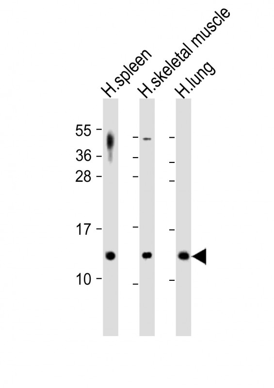
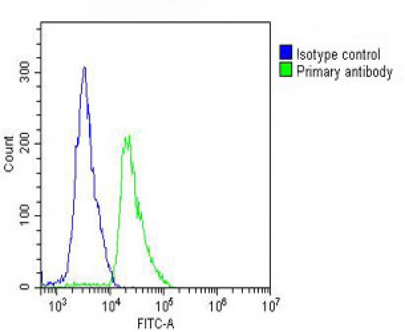
CCL17 Antibody
Purified Rabbit Polyclonal Antibody (Pab)
- SPECIFICATION
- CITATIONS: 1
- PROTOCOLS
- BACKGROUND

Application
| WB, FC, E |
|---|---|
| Primary Accession | Q92583 |
| Reactivity | Human |
| Host | Rabbit |
| Clonality | polyclonal |
| Isotype | Rabbit IgG |
| Calculated MW | 10507 Da |
| Gene ID | 6361 |
|---|---|
| Other Names | C-C motif chemokine 17, CC chemokine TARC, Small-inducible cytokine A17, Thymus and activation-regulated chemokine, CCL17, SCYA17, TARC |
| Target/Specificity | This CCL17 antibody is generated from a rabbit immunized with a recombinant protein of human CCL17. |
| Dilution | WB~~1:2000 FC~~1:25 E~~Use at an assay dependent concentration. |
| Format | Purified polyclonal antibody supplied in PBS with 0.09% (W/V) sodium azide. This antibody is purified through a protein A column, followed by peptide affinity purification. |
| Storage | Maintain refrigerated at 2-8°C for up to 2 weeks. For long term storage store at -20°C in small aliquots to prevent freeze-thaw cycles. |
| Precautions | CCL17 Antibody is for research use only and not for use in diagnostic or therapeutic procedures. |
| Name | CCL17 |
|---|---|
| Synonyms | SCYA17, TARC |
| Function | Chemokine, which displays chemotactic activity for T lymphocytes, preferentially Th2 cells, but not monocytes or granulocytes. Therefore plays an important role in a wide range of inflammatory and immunological processes (PubMed:8702936, PubMed:9169480). Acts by binding to CCR4 at T-cell surface (PubMed:10540332, PubMed:9169480). Mediates GM-CSF/CSF2-driven pain and inflammation (PubMed:27525438). In the brain, required to maintain the typical, highly branched morphology of hippocampal microglia under homeostatic conditions. May be important for the appropriate adaptation of microglial morphology and synaptic plasticity to acute lipopolysaccharide (LPS)-induced neuroinflammation (By similarity). Plays a role in wound healing, mainly by inducing fibroblast migration into the wound (By similarity). |
| Cellular Location | Secreted |
| Tissue Location | Constitutively expressed in thymus. Detected at lower levels in the lung, colon and small intestine (PubMed:8702936) Expressed in stimulated peripheral blood mononuclear cells, but not in resting cells (PubMed:8702936). |

Provided below are standard protocols that you may find useful for product applications.
Background
Chemotactic factor for T-lymphocytes but not monocytes or granulocytes. May play a role in T-cell development in thymus and in trafficking and activation of mature T-cells. Binds to CCR4.
References
Imai T.,et al.J. Biol. Chem. 271:21514-21521(1996).
Livingston R.J.,et al.Submitted (OCT-2006) to the EMBL/GenBank/DDBJ databases.
Loftus B.J.,et al.Genomics 60:295-308(1999).
Mural R.J.,et al.Submitted (JUL-2005) to the EMBL/GenBank/DDBJ databases.
Bernardini G.,et al.Eur. J. Immunol. 28:582-588(1998).
If you have used an Abcepta product and would like to share how it has performed, please click on the "Submit Review" button and provide the requested information. Our staff will examine and post your review and contact you if needed.
If you have any additional inquiries please email technical services at tech@abcepta.com.
Ordering Information
Other Products
Shipping Information
